
Gen Z arriving at work places
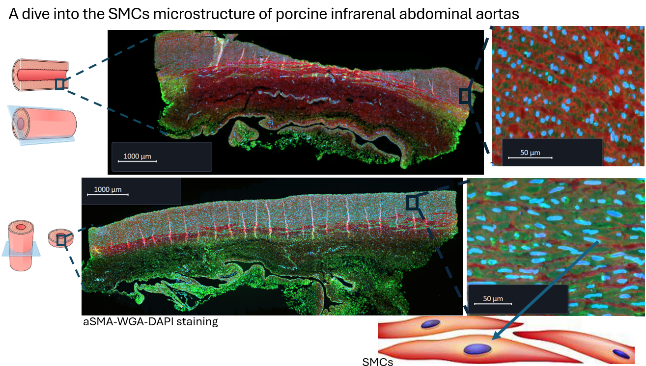
Classical histology in 3D ?
I cannot put my finger on it
Christmas lights
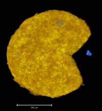
Micro- Pacman
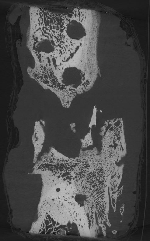
Haunted sample
Aïki noodles
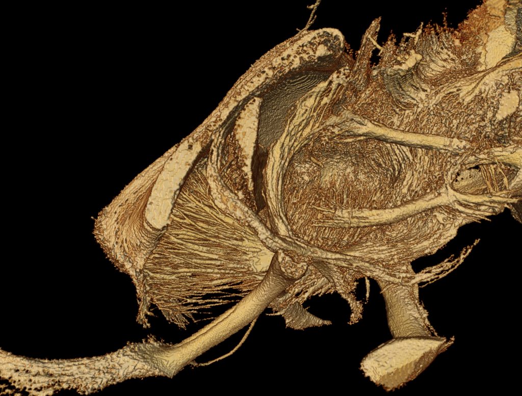
Bony fox race
DIY, not always a good idea
Colorimetric CECT
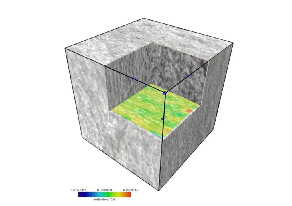
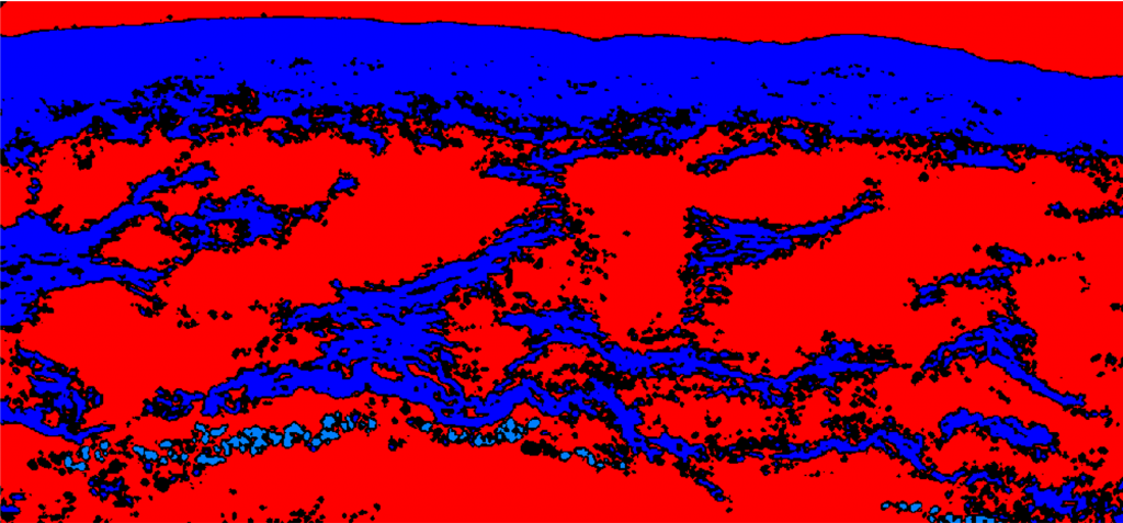
Aorta-shearing
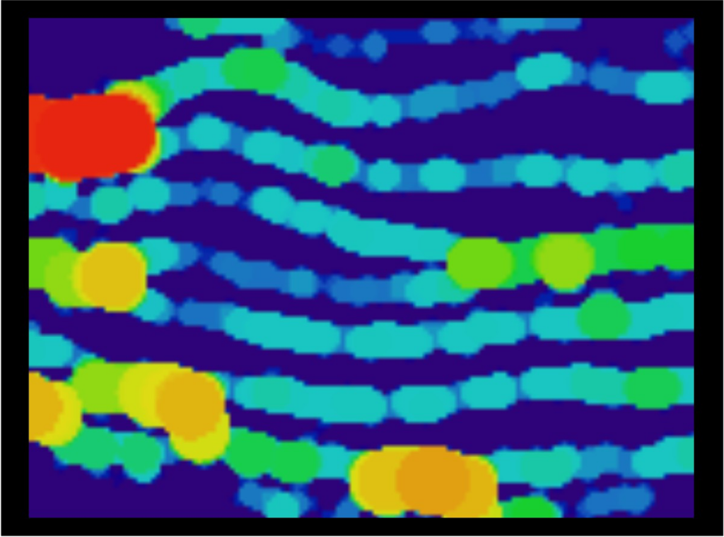
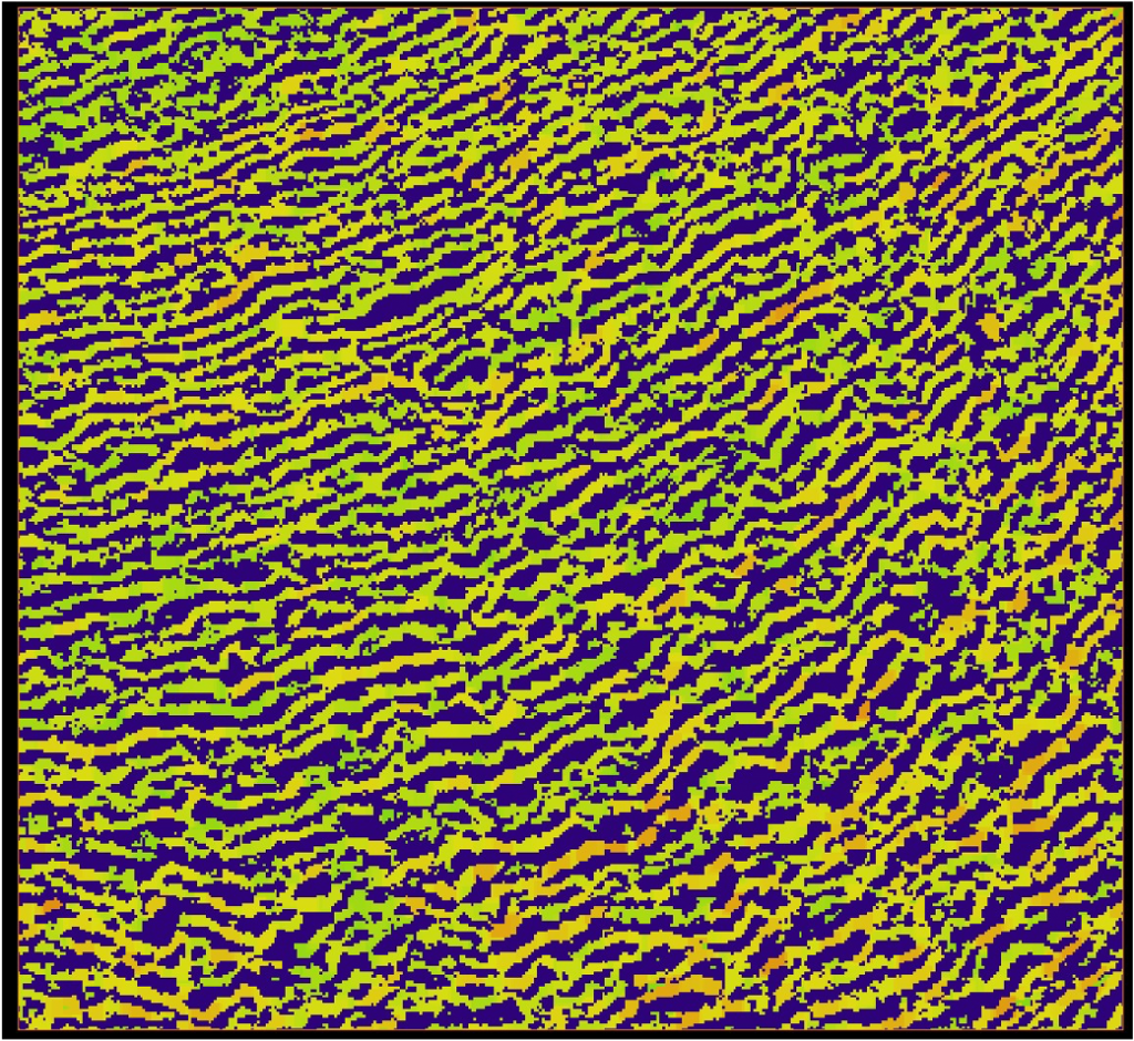
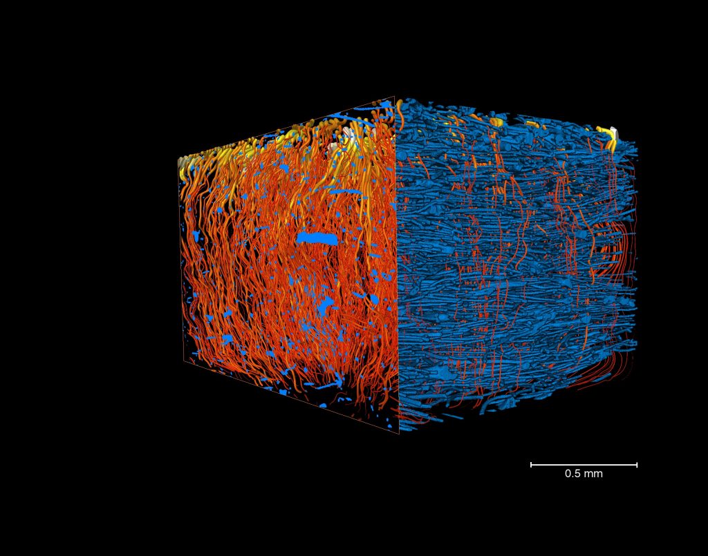
Fatty elastin
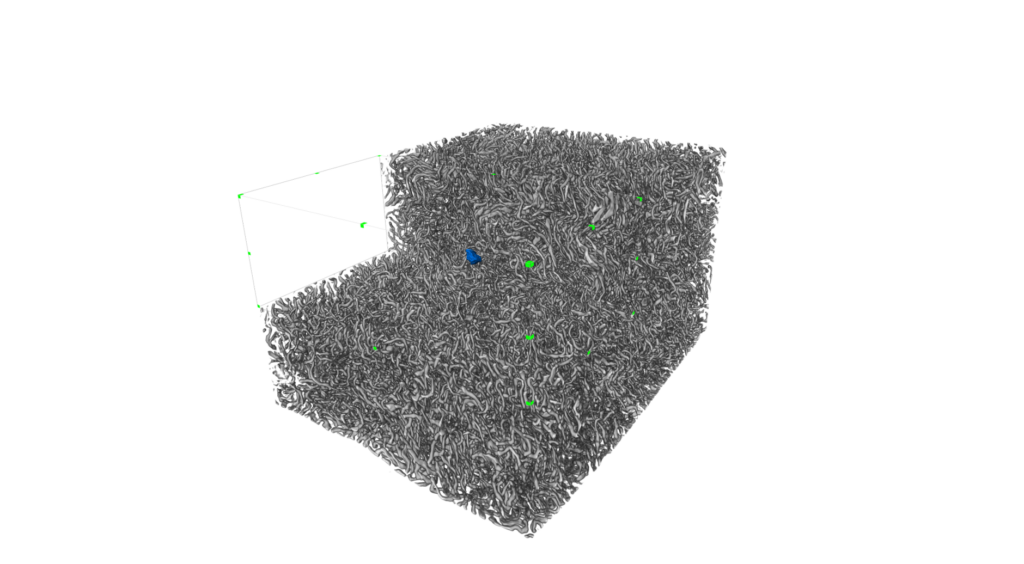
Thickness analysis of the elastin in a rat aorta
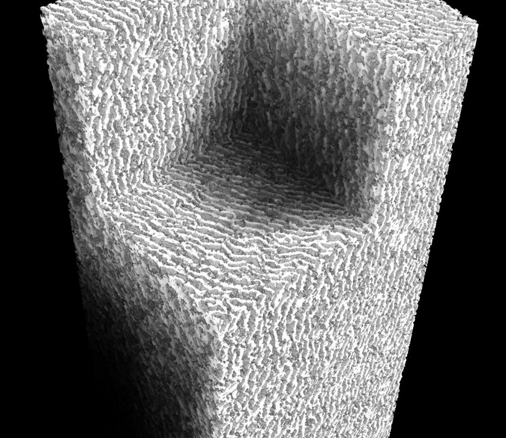
Aortic illusion
Elastic sheets in the media of the porcine aorta
Snakes swimming in iron
Covered by wings, tyramide found its protection
Sauron’s pork (eye)orta
Tornado vs. vessel
Aortas in the jungle
White matter matters
Viscose mop
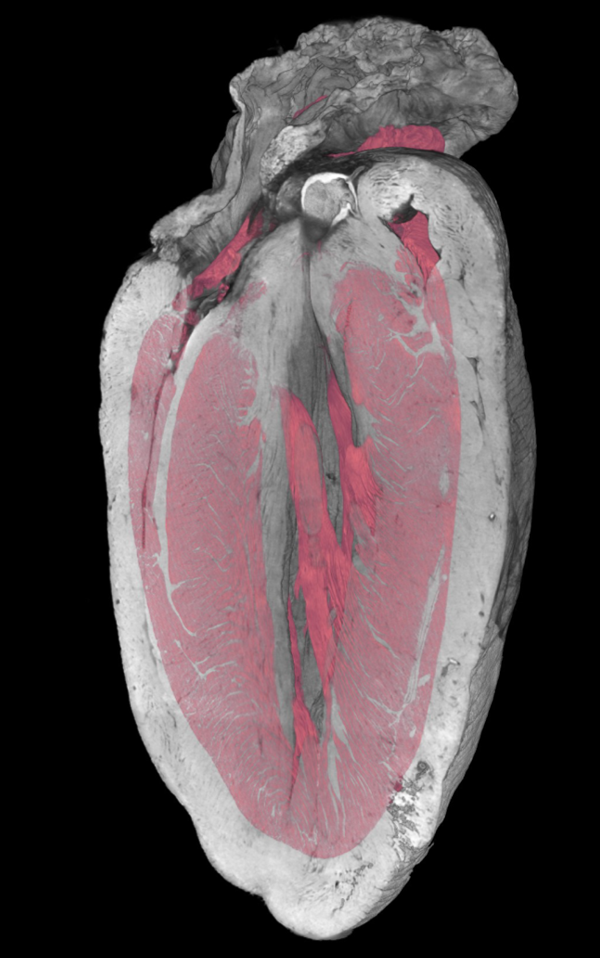
Heartception
Heart stained with Lugol (pink) and Hf-WD POM (grey)
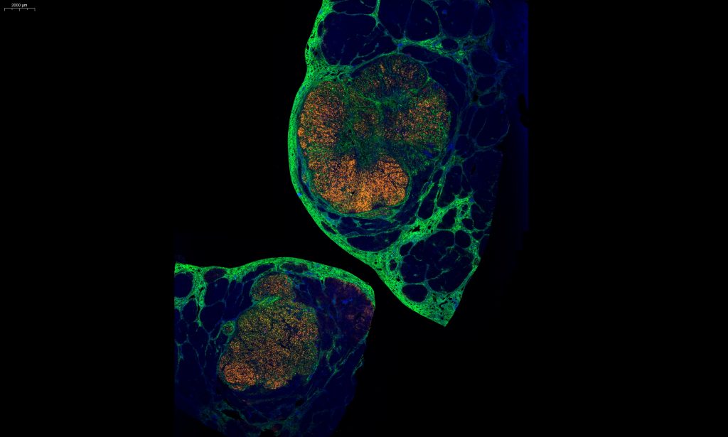
Hepatocarcinoma
Fibrosis and tumor of the liver
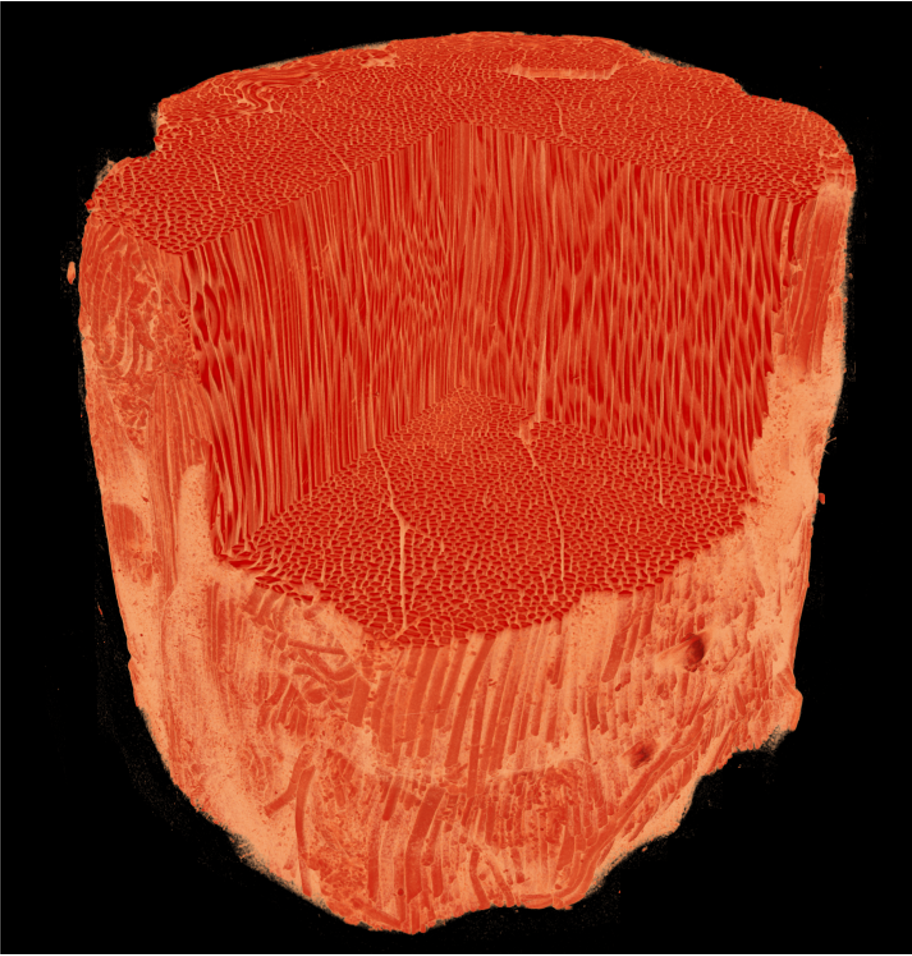
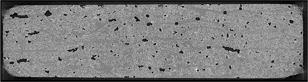
The steaks are high
3D bovine muscle tissue
Staining time is money
Staining of a piece of porcine aorta
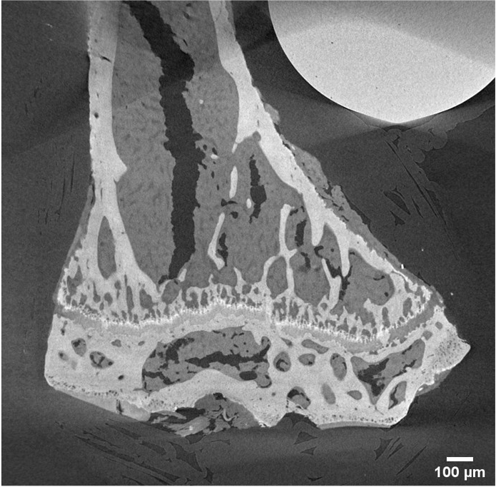
Break a cartileg
Cartilage
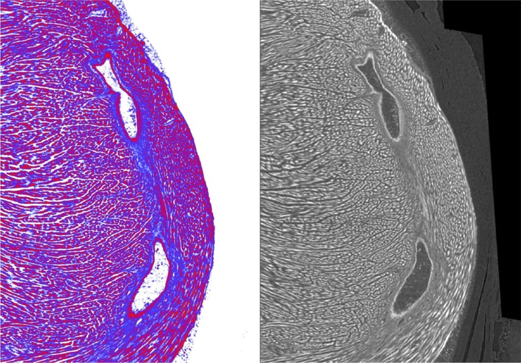
2D or not 2D
Histology – microCT comparison
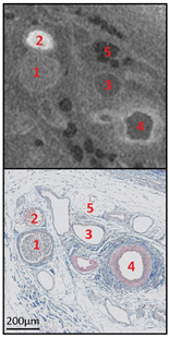
Human auricles stained with Hf-WD POM (CE-CT on top and histology with Masson’s Trichrome on bottom). 1 = nerve, 2 = small vein filled with red blood cells, 3 = small empty vein, 4 = small artery, 5 = adipose tissue
Human ear
Stained with POM, Phoenix Nanotom m, (GE Measurement and Control Solutions, Wunstorf, Germany)
Human calcified heart valve
Stained with POM at 5 µm voxel size, Phoenix Nanotom m, (GE Measurement and Control Solutions, Wunstorf, Germany)
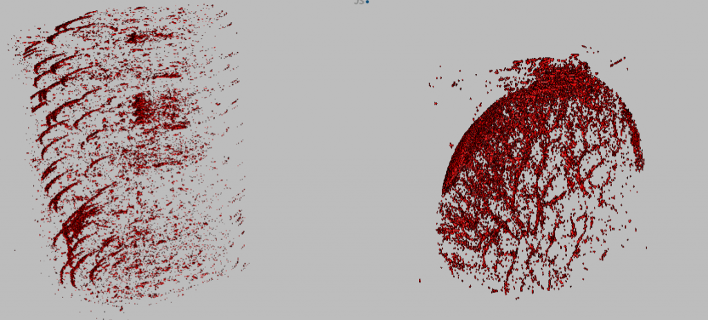
Rat aorta implanted with an iron-based alloy wire
Stained with Hf-WD POM at 1.5 µm voxel size, Phoenix Nanotom m, (GE Measurement and Control Solutions, Wunstorf, Germany)
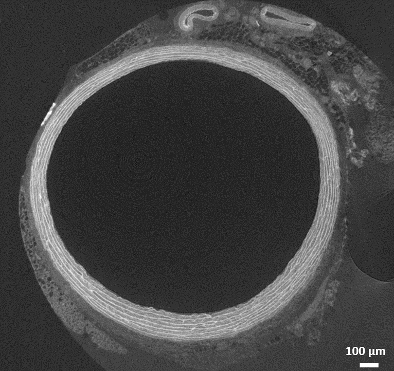
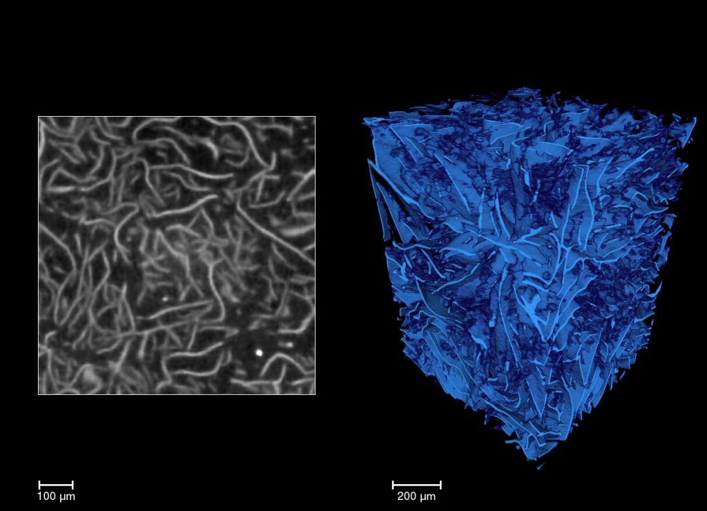
Rat aorta
Stained with Hf-WD POM at 1.5 µm voxel size, Phoenix Nanotom m, (GE Measurement and Control Solutions, Wunstorf, Germany)